Hammer Toe

Overview
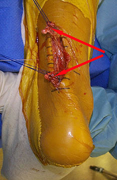
A Hammer Toe is a toe that is bent because of a muscle imbalance around the toe joints. The imbalance causes the toe to bend at one or more joints, pushing the middle of the toe upward in a claw-like position. If you notice such changes, it is important to seek proper treatment. Hammer toes never get better without some type of intervention and the sooner it is treated, the better the outcome.
Causes
While there are a number of causes, there aren't many specific risk factors for hammertoes, women tend to get these problems more than men, but they occur without rhyme or reason. Diabetics, however, are more likely to get a hammertoe if they have underlying nerve damage in the toes and feet.
Symptoms
Common symptoms of hammertoes include pain or irritation of the affected toe when wearing shoes. corns and calluses (a buildup of skin) on the toe, between two toes, or on the ball of the foot. Corns are caused by constant friction against the shoe. They may be soft or hard, depending upon their location. Inflammation, redness, or a burning sensation. Contracture of the toe. In more severe cases of hammertoe, open sores may form.
Diagnosis
A hammertoe is usually diagnosed with a physical inspection of your toe. Imaging tests, such as X-rays, may be ordered if you have had a bone, muscle, or ligament injury in your toe.
Non Surgical Treatment
The most common treatment is to wear more comfortable shoes. When choosing a shoe, make sure the toe area is high and broad and has enough room for hammer toes. If there is chronic pain, surgery may be needed to correct a malalignment. Surgical treatments are aimed at loosening up the contracted toe joints to allow them to align properly. Other types of treatment are products designed to relieve hammer toes, such as hammer toe crests and hammer toe splints. These devices will help hold down the hammer toe and provide relief to the forefoot. Gel toe shields and gel toe caps can also be used. Gel toe shields and toe caps will help eliminate friction between the shoe and the toe, while providing comfort and lubrication.
Surgical Treatment
In advanced cases in which the toe has become stiff and permanently bent, the toe can be straightened with surgery. One type of surgery involves removing a small section of the toe bone to allow the toe to lie flat. Surgery for hammertoe usually is classified as a cosmetic procedure. Cosmetic foot surgeries sometimes result in complications such as pain or numbness, so it's better to treat the problem with a shoe that fits properly.
Prevention
Have your feet properly measured, make sure that, while standing, there is a centimetre (? thumb) of space for your longest toe at the end of each shoe. Buy shoes that fit the longer foot. Shop at the end of the day, when foot swelling is greatest. Don't go by numbers, sizes vary by brand, so make certain your shoes are comfortable. Wear wide shoes with resilient soles, avoid shoes with pointed toes.

RosellaHannafin 發表在 痞客邦 留言(0) 人氣()

Pain In Arch

Overview
Your foot has an incredible design. All of the parts fit together in such a way as to be flexible and moveable while still being very strong. One of the important parts of the foot is your arch. Located in the middle of your foot, it is slightly raised off the ground and helps you absorb the impact of every step. Sometimes, however, it can ache and make walking or standing around very uncomfortable.
Causes
Stress fractures, plantar fasciitis, and acute and chronic arthritis are most commonly the result of repetitive micro-trauma injuries. Micro-trauma injuries are caused when the structures of the body are stressed and re-stressed to the point that damage occurs in the tissues. Factors that commonly contribute to this injury can be running on uneven surfaces or surfaces that are too hard or too soft, shoes that have poor force-absorption qualities, or going too hard or too long during repeated exercise bouts.
Symptoms
The primary symptom is pain or aching in the arch area. This can be accompanied by inflammation and tenderness. If the pain is caused by the plantar fascia, it is likely to be considerably more severe in the mornings due to the muscles being unused. If you notice that the twinges of pain you have are most commonly associated with or immediately after exercise, you might want to visit a good athletic shoe store to make sure you are wearing the right kind of shoe. Another cause of arch pain is plantar fasciitis. The plantar fascia is a tough tissue structure that holds the bottom part of the arch in place. The fascia often becomes inflamed and sore, usually as a result of repetitive motion (for example, very common in those who stand on their feet for work). The pain is often noticeable first thing in the morning and worse with activity.
Diagnosis
Flat feet are easy to identify while standing or walking. When someone with flat feet stands, their inner foot or arch flattens and their foot may roll over to the inner side. This is known as overpronation. To see whether your foot overpronates, stand on tiptoes or push your big toe back as far as possible. If the arch of your foot doesn't appear, your foot is likely to overpronate when you walk or run. It can be difficult to tell whether a child has flat feet because their arches may not fully develop until they're 10 years of age.
Non Surgical Treatment
Tight arches, overpronation and flat feet as well as tight calves all lead to inflammation of the connective tissue that forms this arch of your foot. This condition, plantar fasciitis, can be very painful, from your first step in the morning. Fortunately, treatment is easier than most. No operations, no fancy pulsed waves, no night splints necessary. Good arch supports? full length flexible (not hard) orthotics are key. Stretches of the calf relieve arch tightness as the heel bone acts like a fulcrum pulling back the arch tissues if too tight. Regularly perform the gastroc and soleus stretches that you learned at the gym. If lazy, buy "Pro-Stretch" on line which helps you stretch your calves easily. Use it often. You can't stretch too often, only too little. Finally, the magic cure is to roll a golf ball under the arch for half-hour once a day. (I know; a half-hour is a long time to perform one exercise, but that's what it takes. Once you master this exercise, it is easy to do while you work at your desk or are watching a half hour TV program.) This may hurt the first week. Keep going because by week two, after you go over the pain hump, the pain will be gone.
Surgical Treatment
Foot surgery is difficult, especially when large amounts of deformity correction are needed. The ability to bring the foot into a new position may not be lasting, even if everything looks perfect in the operating room. The goal is to provide improved position and function of the foot and ankle. In some patients with very severe deformity, the goal is a foot that functions well in a brace. There are complications that relate to surgery in general. These include the risks associated with anesthesia, infection, damage to nerves and blood vessels, and bleeding or blood clots. Particular complications associated with cavus foot surgery include incomplete correction of deformity, return of deformity and incomplete fusion.
Stretching Exercises
Ankle evert or strengthening. Lie on your side with your feet hanging off the end of your bed or a weight bench. Bend the toes of the foot that is closer to the ceiling slightly toward your head. This is the starting position. Now raise your toes toward the ceiling while keeping the rest of your leg stationary. Return to the starting position. Reps. 10-15. Now point your toes slightly away from your head. This is the starting position. Raise your toes toward the ceiling. Return to the starting position. Reps. 10-15. Ankle invertor strengthening. Same as above, but do the exercises with the foot that is closer to the floor. Dorsiflexor strengthening. Sit on a desk, table, or counter so that your feet don?t touch the ground. Let your feet dangle comfortably. Bend your foot upward as far as you can comfortably go. Do not let your foot pull inward or outward. Return to the starting position. Reps. 10-15.

RosellaHannafin 發表在 痞客邦 留言(0) 人氣()

Foot Arch Pain

Overview
Flexible flatfeet are considered normal in young children because babies are not born with a normal arch. The arch may not form fully until sometime between ages 7 and 10. Even in adulthood, 15% to 25% of people have flexible flatfeet. Most of these people never develop symptoms. In many adults who have had flexible flatfeet since childhood, the missing arch is an inherited condition related to a general looseness of ligaments. These people usually have extremely flexible, very mobile joints throughout the body, not only in the feet. Flatfeet also can develop during adulthood. Causes include joint disease, such as rheumatoid arthritis, and disorders of nerve function (neuropathy).
Causes
Arch and heel pain is usually the result of faulty biomechanics (walking gait abnormalities) that place too much stress on the heel bone and the soft tissues that attach to it. This increased stress causes local inflammation and pain. The most common cause of the stress is a condition where the inside arch of the foot flattens more than it should (often known as "over-pronation"). When the arch of the foot flattens, it also gets longer, causing a stretch on the plantar fascia. In response, the heel becomes inflamed where the plantar fascia attaches.
Symptoms
Arch pain may have a variety of different causes. Proper evaluation and diagnosis of arch pain is essential in planning treatment. A good general guideline is to compare the injured side to the uninjured side. Injury may present itself as a distinguishable lump, a gap felt at that location, or a "crunchy" feeling on that spot caused by inflammation. The type, causes, and severity of pain are also good indicators of the severity of the injury.
Diagnosis
In people with flat feet, the instep of the foot comes in contact with the ground when standing. To diagnose the problem, the health care provider will ask you to stand on your toes. If an arch forms,the flat foot is called flexible. You will not need any more tests or treatment. If the arch does not form with toe-standing (called rigid flat feet), or if there is pain, other tests may be needed, including a CT scan to look at the bones in the foot. MRI scan to look at the tendons in the foot. X-ray of the foot.
Non Surgical Treatment
The most effective treatment for foot arch pain and strain is to use an arch support. The arch support sits under the foot and stops the arch of the foot from collapsing, thereby preventing the stretch of the arch pad which causes pain and discomfort. Wearing an arch support in slippers or house shoes can also prevent pain in the mornings when discomfort it most common and severe. Arch supports usually relieve symptoms within a few days.
Surgical Treatment
In cases where cast immobilization, orthoses and shoe therapy have failed, surgery is the next alternative. The goal of surgery and non-surgical treatment is to eliminate pain, stop progression of the deformity and improve mobility of the patient. Opinions vary as to the best surgical treatment for adult acquired flatfoot. Procedures commonly used to correct the condition include tendon debridement, tendon transfers, osteotomies (cutting and repositioning of bone) and joint fusions.
Prevention
Stretch and strengthen important muscles in your feet, ankles and legs in order to guard against future strain. Make sure to acquire suitable arch supports and inserts if necessary, and that your shoes are shock absorbent and in good condition. Wearing tattered shoes provides no protection, and runners should replace their footwear before exceeding 500 miles of usage. Athletes new to arch supports should gradually build their training routine, allowing their feet to become accustomed to a new stance.
Stretching Exercises
Achilles stretch. Stand with the ball of one foot on a stair. Reach for the step below with your heel until you feel a stretch in the arch of your foot. Hold this position for 15 to 30 seconds and then relax. Repeat 3 times. Balance and reach exercises. Stand next to a chair with your injured leg farther from the chair. The chair will provide support if you need it. Stand on the foot of your injured leg and bend your knee slightly. Try to raise the arch of this foot while keeping your big toe on the floor. Keep your foot in this position. With the hand that is farther away from the chair, reach forward in front of you by bending at the waist. Avoid bending your knee any more as you do this. Repeat this 15 times. To make the exercise more challenging, reach farther in front of you. Do 2 sets of 15. While keeping your arch raised, reach the hand that is farther away from the chair across your body toward the chair. The farther you reach, the more challenging the exercise. Do 2 sets of 15. Towel pickup. With your heel on the ground, pick up a towel with your toes. Release. Repeat 10 to 20 times. When this gets easy, add more resistance by placing a book or small weight on the towel. Resisted ankle plantar flexion. Sit with your injured leg stretched out in front of you. Loop the tubing around the ball of your foot. Hold the ends of the tubing with both hands. Gently press the ball of your foot down and point your toes, stretching the tubing. Return to the starting position. Do 2 sets of 15. Resisted ankle dorsiflexion. Tie a knot in one end of the elastic tubing and shut the knot in a door. Tie a loop in the other end of the tubing and put the foot on your injured side through the loop so that the tubing goes around the top of the foot. Sit facing the door with your injured leg straight out in front of you. Move away from the door until there is tension in the tubing. Keeping your leg straight, pull the top of your foot toward your body, stretching the tubing. Slowly return to the starting position. Do 2 sets of 15. Heel raise. Stand behind a chair or counter with both feet flat on the floor. Using the chair or counter as a support, rise up onto your toes and hold for 5 seconds. Then slowly lower yourself down without holding onto the support. (It's OK to keep holding onto the support if you need to.) When this exercise becomes less painful, try doing this exercise while you are standing on the injured leg only. Repeat 15 times. Do 2 sets of 15. Rest 30 seconds between sets.

RosellaHannafin 發表在 痞客邦 留言(0) 人氣()

+lt`;'(G`HF3 Adult Aquired Flat Foot.txtY!JəT]ΙY3&IJH4ZE&sECi'#SY:76yMfbG,_H ^]@dA$ן}_ϧhW\r`ϋy?vbywKk^trf,1z("[ލa;-ǃGXJ| ;};?ϓXHBC_)/>:h`r@:&<o#ƽCË0ogJ4V=qtNj?tE!ͬx5ɋEapϗ[;ls3Lpw@iNpjd 5v[K8]/L4ؾx[]R}w.8'ik(NgǶ}wۇ 9/gnG(2P} Jp%1ÛFW[~8 E;dM}υnNG@g8fy/t(jwϊM׫r,^7Aνcɤ\>(sC&w=kGӳtr|v
(B)ϕ:fw_ ꇮB>la>-=5᧼tTm=@d<0sl>9X .y3G,v_9q(lqv:F l}S~o#+vR0ۏ@"臻TMrX>9߽|w./3ۧVB|yCb`+l?UC/\qXg>MVxϫƏPqIoݯB87N7ǧbbz/_"C <+ KCw*R&ԗWܸ)*AU
rx~ހҙό\}F'q莇ǿ@dzau
ֶ=Z"uQjk3=J
@Ǟ|72BǪcZMl"@7ziGG!)4<Q&i*w0|<n
ZW_nMa: 6c!ش
AKyj,#MdȲfy<]LygVWL`Q
L_eP
p=Gj|TIs@6!'P(fhH=*b+\ǴR:&<?<M`(NǒJj}OEc?m'@χSb?`;2A((A +3E1pP7YTK;kVCV$(Gn20829<OWg|zjU;ZҐix
TᓸTm1Ҿ?XID7
yZمv!2V" C3RF2 i
&͙VֹvD4& G/Ibeu*S0O0N#74vfnҔ
hqh5qYfmbS]q
%sw%-d׎]QT"zo|y9j&xʻR4qig t
CF#A֛Nஔק>(@f7MXQ^@$ɋLi
"PpE!ucLNMt1n-9}Zii#HH(2S;PhBt@wj4K (%`JaЕZj1[HTq4wSZSݕ(W$z,>& ؙQXR\ۃ:JE)K9ߑY*/:ۛi"9
HWHˎ$<HXB5w8R*iLHl[PZwc4]AWD
E2b睖!p7d67<Hf<o.ebpĸxPŕ[m]q[.Qk0XG ٌzHΒfou'gFMC UgX#P)ݱ];B;RftO^J%)\dUXa%ySz?;gր慚@oU=1'5,Yh ]i^1ֈKZ~BP'|6MExG2c'$'[';baKzA4ΕFR>fi09tÎ87/4'.GV#NiBF)?>bFDo%O5drҾ'ի[7T^Yg[n*dEᓛQ]^Q-Jݕ,vYoQ-rt'#jί;?> UWfތyޢq*u>pZ@o>2_~\8uy=½K#ժY甶21CQG@E"AZ)hw-lz<+usA0*)v^wu7|ժw-o^IOWӟ~X6<`6ZlQ9~wޯ¯.Uȿn>K:U[rz6<֩EZ>?WWߎCC;Uf߫鱛b%t`۱Չm8C15?g¿;ǫVo<V%.N<߅o7W#CWOc+)+V\gE^oh=
8[KdZ!?W ZLQCy߫ZۚGnf1H
!;A?f'j?iw1l~0aD8l=oRikd;7~P+`͓\~y~7mvÜ>_o罟+x9 O5?o;olv*ծo
>pʿ.~̿jīZ~\J#8h8M-o(bq%4ۯAwA%1?Ό$v.EMOXEZ6T׫֌z|
b
J6<`,_ҩ/nՊUWVի@^]k%5I p_l}0hN^z``ǒ0tNrU5%a׀{b2tv*5mINfFxY**O=1@cKmfi9r!D+u*ٱSaECOA
1ttog_Mh=1K ó
B\,?FWVXJ,V_.T-ѐuXF-fm@dԅj4=vvvH`Kx30#⋫2IW9x4HS
kwEX?B5#%V(pVkXBc`i6!N0o|5488mYzu^~q^{,Xgh%5io:-3
ZE<7^==bgb[]nlF]D=kc2W=^مP<ݹt>RҥTKEb*!28v:5*=C]jE܁j`-]4u~ P^ū?>TՃŋ#GG韤v?Y갗FC=I1?8V58,Χu%!ce@5V^͠gV+Xn<z%GJѱu2Fm6l.Fī6kV8+Y|W~^ԫ8e_!*R)cr5_!bUr~܍WS/O!O(;,Y+V^Rbeq*[nS\:‘!Je$u'@w(;NnV8ix_Vj+P&Z̳ndv_t+ұ*tlY"*+Zq܉xyO;wх%d1d:^'5жdie5odɿFWZq\2skU+ښjn lk eסys=>
2okjlC2@m?(]c$y$% s͏[ϳ&l.tLih?'D8a?f
pá-yG.e,
J?;)Q~xO@/{Yz\@O<A!K傖)]L`Ǐќ`/W4 ]~^:\<5o?I܅IC/0xM%Ӄ?f>(Oy`E! [v-ZV+W8\@?h0>]gh
nYplE|N2N
Et^.
pfO{ UW{_TCp490)1m>&2\a͞CR^e"ϯJzR媥:\U@Vd=zZ<wX>OH
W~stV~k
|
叇86)cr
|(_
_w/j|ڡ,uuQpjD!n'WuwH2^pBYpjm9ɇ!l+T3-d{S a&L=,>ǃz#GH~UܬE䏊X[lKPZW=M٫~n;w۟5߼,82u3ϋV\~զф?4 W./O7,2̔[d *zW-Va?30>DۂUizfv5^d
-رkhqV/AqW&2{=
ԟ*\d,LR§=:xlWjK@79s
{dzcr@AqD5oJelثY9Malr*ȤWVkR*]$UɚȬo9wjKe9Ƨ=mԤ<`Rjo29';O;'&l2R,O~ߏH[Y
LejV
CW%"+G鞱SHH ڿiSô2Rϓ-~xó5WsU{-DK{Kع`Q\U^<3RԘ$k͕!8C-&Rk:l7({{cR| ُE"ݦPs$I#sIzQJ[ lЗ/Ks쓍U6<0`\.ٶ"vdqQ`ԫ:Hl8UZD0wp.='Z
7)R/xڼvq4ېfX~i$R?4Mh$p#vhgC#mr
4)Vذ%\ls S,xD59>eQΑ%C+e_^ŹU-RIZ;.D5%l6([%)%T
ʒ,<&9ݏzXoit' DLVXaS֦0hnMTOM™%Ǎ/Ε!4#W{lXa-Gt6_wi
r(.^ɨ=SaG,SĩBȩGPsoldo4'
Ȱ+m66i俲*XrEEɲS.7ZoV.Sig<5pQN'A%wfʲ3F;
">#[ɁUI2soIY)9"oD3!丫i7f*UKjg|EGiFU/R_K&*vy1/ߚv!ʾfI"UCDťO WQ~$
S98A1a6%7k\mmDlH(&DgBضGw)6(Q7&wiT$J%.~I]jv

RosellaHannafin 發表在 痞客邦 留言(0) 人氣()

Achilles Tendon

Overview
Achilles Tendon Rupture Surgery is a surgical procedure is used to repair a ruptured Achilles tendon. There are two types of surgery an open surgery and a percutaneous surgery, in both cases the surgeon sews the tendon back together through the incision helping the tendon to heal properly and restore function to the foot and ankle. If the injury or rupture has caused severe swelling the surgery may be delayed for a short period of time to allow the swelling to go down.
Causes
The tendon usually ruptures without any warning. It is most common in men between the ages of 40-50, who play sports intermittently, such as badminton and squash. There was probably some degeneration in the tendon before the rupture which may or may not have been causing symptoms.
Symptoms
Symptoms of an Achilles tendon rupture include sensation that someone or something has hit the back of the calf muscle, sudden pain, pain when walking, weakness in the leg, which is particularly noticeable when trying to push off while walking and there is not sufficient strength to do so.
Diagnosis
To diagnose an Achilles tendon injury, your health care provider will give you a thorough physical exam. He or she may want to see you walk or run to look for problems that might have contributed to your Achilles tendon injury.
Non Surgical Treatment
This condition should be diagnosed and treated as soon as possible, because prompt treatment probably improves recovery. You may need to be referred urgently to see a doctor in an orthopaedic department or accident and emergency department. Meanwhile, if a ruptured Achilles tendon is suspected, you should not put any weight on that foot, so do not walk on it at all.Treatment options for an Achilles tendon rupture include surgical and non-surgical approaches. The decision of whether to proceed with surgery or non-surgical treatment is based on the severity of the rupture and the patient?s health status and activity level. Non-surgical treatment, which is generally associated with a higher rate of re-rupture, is selected for minor ruptures, less active patients, and those with medical conditions that prevent them from undergoing surgery. Non-surgical treatment involves use of a cast, walking boot, or brace to restrict motion and allow the torn tendon to heal.
Surgical Treatment
Operative treatment involves a 6cm incision along the inner side of the tendon. The torn ends are then strongly stitched together with the correct tension. After the operation a below knee half cast is applied for 2 weeks. At 2 weeks a brace will be applied that will allow you to move the foot and fully weight-bear for a further 6 weeks. After this you will need physiotherapy. Surgery carries the general risks of any operation but the risk of re-rupture is greatly reduced to 2%. The best form of treatment is controversial with good results being obtained by both methods but surgery is generally recommended for patients under 60 years of age who are fit and active with an intra-substance tear.
Prevention
To reduce your chance of developing Achilles tendon problems, follow the following tips. Stretch and strengthen calf muscles. Stretch your calf to the point at which you feel a noticeable pull but not pain. Don't bounce during a stretch. Calf-strengthening exercises can also help the muscle and tendon absorb more force and prevent injury. Vary your exercises. Alternate high-impact sports, such as running, with low-impact sports, such as walking, biking or swimming. Avoid activities that place excessive stress on your Achilles tendons, such as hill running and jumping activities. Choose running surfaces carefully. Avoid or limit running on hard or slippery surfaces. Dress properly for cold-weather training and wear well-fitting athletic shoes with proper cushioning in the heels. Increase training intensity slowly. Achilles tendon injuries commonly occur after abruptly increasing training intensity. Increase the distance, duration and frequency of your training by no more than 10 percent each week.

RosellaHannafin 發表在 痞客邦 留言(0) 人氣()

Achilles Tendon

Overview
A rupture of the Achilles tendon means that there has been either a complete, or partial, tear of the tendon which connects the calf muscles to the heel bone. Usually this occurs just above insertion on the heel bone, although it can happen anywhere along the course of the tendon. Achilles tendon rupture occurs in people that engage in strenuous activity, who are usually sedentary and have weakened tendons, or in people who have had previous chronic injury to their Achilles tendons. Previous injury to the tendon can be caused by overuse, improper stretching habits, worn-out or improperly fitting shoes, or poor biomechanics (flat-feet). The risk of tendon rupture is also increased with the use of quinolone antibiotics (e.g. ciprofloxacin, Levaquin).
Causes
People who commonly fall victim to Achilles rupture or tear include recreational athletes, people of old age, individuals with previous Achilles tendon tears or ruptures, previous tendon injections or quinolone use, extreme changes in training intensity or activity level, and participation in a new activity. Most cases of Achilles tendon rupture are traumatic sports injuries. The average age of patients is 29-40 years with a male-to-female ratio of nearly 20:1. Fluoroquinolone antibiotics, such as ciprofloxacin, and glucocorticoids have been linked with an increased risk of Achilles tendon rupture. Direct steroid injections into the tendon have also been linked to rupture. Quinolone has been associated with Achilles tendinitis and Achilles tendon ruptures for some time. Quinolones are antibacterial agents that act at the level of DNA by inhibiting DNA Gyrase. DNA Gyrase is an enzyme used to unwind double stranded DNA which is essential to DNA Replication. Quinolone is specialized in the fact that it can attack bacterial DNA and prevent them from replicating by this process, and are frequently prescribed to the elderly. Approximately 2% to 6% of all elderly people over the age of 60 who have had Achilles ruptures can be attributed to the use of quinolones.
Symptoms
Symptoms of an Achilles tendon rupture usually directly follow a traumatic event where the foot is forced in an upward position at the ankle, causing a sudden tight stretch of the Achilles tendon. There can also be a direct blow to the tendon causing a rupture. There is typically a popping feeling or even a popping sound described during the occurance of the rupture. Typically there is pain with swelling in the region. Often the patient is unable to put weight on this foot as there is too much pain.
Diagnosis
Laboratory studies usually are not necessary in evaluating and diagnosing an Achilles tendon rupture or injury, although evaluation may help to rule out some of the other possibilities in the differential diagnosis. Plain radiography. Radiographs are more useful for ruling out other injuries than for ruling in Achilles tendon ruptures. Ultrasonography of the leg and thigh can help to evaluate the possibility of deep venous thrombosis and also can be used to rule out a Baker cyst, in experienced hands, ultrasonography can identify a ruptured Achilles tendon or the signs of tendinosis. Magnetic resonance imaging (MRI). MRI can facilitate definitive diagnosis of a disrupted tendon and can be used to distinguish between paratenonitis, tendinosis, and bursitis.
Non Surgical Treatment
Initial treatment for sprains and strains should occur as soon as possible. Remember RICE! Rest the injured part. Pain is the body's signal to not move an injury. Ice the injury. This will limit the swelling and help with the spasm. Compress the injured area. This again, limits the swelling. Be careful not to apply a wrap so tightly that it might act as a tourniquet and cut off the blood supply. Elevate the injured part. This lets gravity help reduce the swelling by allowing fluid and blood to drain downhill to the heart. Over-the-counter pain medication is an option. Acetaminophen (Tylenol) is helpful for pain, but ibuprofen (Motrin, Advil, Nuprin) might be better, because these medications relieve both pain and inflammation. Remember to follow the guidelines on the bottle for appropriate amounts of medicine, especially for children and teens.
Surgical Treatment
An Achilles tendon rupture is a complete tear of the fibrous tissue that connects the heel to the calf muscle. This is often caused by a sudden movement that overextends the tendon and usually occurs while running or playing sports such as basketball or racquetball. Achilles tendon rupture can affect anyone, but occurs most often in middle-aged men.
Prevention
To help reduce your chance of getting Achilles tendon rupture, take the following steps. Do warm-up exercises before an activity and cool down exercises after an activity. Wear proper footwear. Maintain a healthy weight. Rest if you feel pain during an activity. Change your routine. Switch between high-impact activities and low-impact activities. Strengthen your calf muscle with exercises.

RosellaHannafin 發表在 痞客邦 留言(0) 人氣()

1
Blog Stats
⚠️

成人內容提醒

本部落格內容僅限年滿十八歲者瀏覽。
若您未滿十八歲,請立即離開。

已滿十八歲者,亦請勿將內容提供給未成年人士。